ACL tears are one of the most common knee injuries, sidelining more than 100,000 Americans every year. Traditionally, repairing a torn ACL (Anterior Cruciate Ligament) required surgical reconstruction using tissue from another part of the patient’s body or donor tissue. While often effective, this procedure is invasive and requires a lengthy recovery time. But now, thanks to advancements in orthopedic surgery, patients suffering from a torn ACL have an alternative called Bridge Enhanced ACL Restoration, also known as BEAR.
What is the BEAR Procedure?
BEAR is a breakthrough in ACL repair that enables the body to naturally heal on its own. Because synovial fluid, which reduces friction in the knee, prevents formation of blood clots that are necessary for healing, the ACL does not have the ability to heal itself. In the BEAR procedure, instead of removing the damaged ACL and replacing it with donor tissue, a special implant is used to bridge the torn ends of the ligament. This creates an ideal healing environment, enabling the ACL to heal, typically within eight weeks.
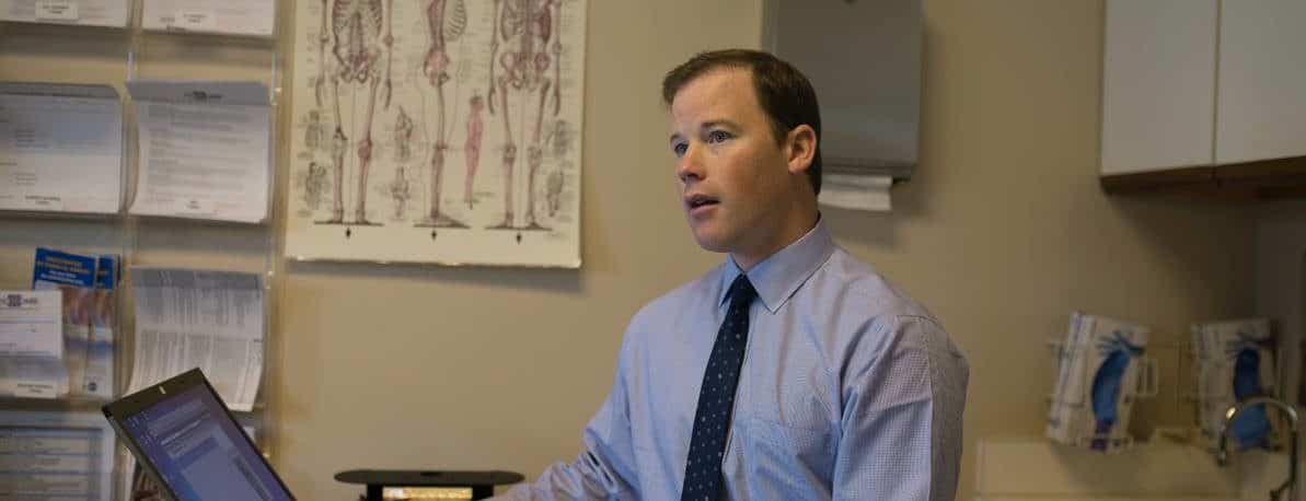
Meet Dr. Welch: Maine’s First BEAR Surgeon
At Atlantic Orthopaedics & Sports Medicine, we’re proud to have Dr. Welch, a pioneer in ACL repair, on our team. Dr. Welch was the first surgeon in Maine to perform the BEAR procedure and remains one of the few specialists in the region to offer it. He performs the surgery at York Hospital and at the New England Center for Orthopedic Surgery in Portsmouth, providing patients with a cutting-edge alternative to traditional ACL reconstruction.
Why Choose the BEAR Procedure?
For many patients, BEAR offers significant advantages over traditional ACL surgery:
- Preserves your own tissue: unlike reconstruction, which requires a graft from another part of the body or a donor, BEAR allows your ACL to heal naturally.
- Better post-operative knee function: because the original ACL is preserved, patients tend to experience less pain and less muscle weakness after surgery.
- Less invasive: the surgery is an outpatient procedure, and does not require harvesting tissue from another part of the body
- No donor tissue risks: There’s no concern over donor graft quality
- Faster healing timeline
Real Results: A Patient’s Journey Back to Running
Patients who undergo the BEAR procedure are seeing amazing outcomes, including Rachel, who recently shared her experience:
“Thanks to Dr. Welch at Atlantic Orthopaedics & Sports Medicine for the amazing BEAR procedure and Shantelle and Ian at Coppola Physical Therapy for helping me get back on the road as quickly as I did. I’m so lucky to have this team and my family and friends cheering me on.”
Six months after her surgery, Rachel was back to running–something that once felt impossible. Her success story is just one of many that demonstrate the life-changing potential of this innovative ACL repair method.
Who is Eligible for BEAR?
The BEAR implant was cleared by the FDA for patients who meet the following criteria:
- Must be at least 14 years old.
- Must have a complete ACL rupture, as confirmed by MRI.
- Must have an ACL stump attached to the tibia.
The team at Atlantic Orthopaedics & Sports Medicine are committed to bringing the latest advancements in orthopedic care to our patients. Dr. Welch continues to be at the forefront of ACL treatment, helping patients regain knee strength and mobility with a less invasive approach than traditional reconstruction. If you’ve suffered an ACL tear and are exploring your options, the BEAR procedure may be the right choice for you. Contact Atlantic Orthopaedics today to schedule a consultation with Dr. Welch and learn more.