Is it Too Late for Joint Replacement Surgery? 

When is joint replacement surgery called for? 

Patients suffering from chronic knee or hip pain know that it can seriously impact mobility as well as overall quality of life. In addition to pain, symptoms may include swelling, stiffness, muscle weakness, and limited range of motion. Causes range from rheumatoid or osteoarthritis to trauma to repetitive strain on the joint. After diagnosis, non-surgical treatments like anti-inflammatory medications, physical therapy, and injections are the first course of action when treating persistent joint pain, but these methods aren’t always effective.  

What is involved in joint replacement surgery? 

The surgical screening process begins with a full clinical evaluation, using advanced imaging to determine the extent of the joint damage. The orthopedic surgeon will make an incision to access the damaged bone, tendon, and tissue, and replace the knee or hip joint with a prosthetic made of plastic, metal, or ceramic. The procedure can be done using a conventional approach or with robotic-assistance. The physicians at Atlantic Orthopaedics are at the forefront of innovation in knee and hip replacement techniques and technology and have pioneered the use of minimally-invasive robotic-assisted surgery.  

What can I expect after surgery? 

Commitment to post-operative care is a key factor in long term success. Recovery time will vary based on the individual patient’s response to the procedure and the surgical approach used. Proper recovery usually includes daily movement of the affected joint, physical therapy, and wound care management, along with a post-op follow up with your surgeon.  

What if I’ve waited too long to be eligible for joint replacement surgery? 

Because damage to cartilage, ligaments, and bone is progressive and won’t improve on its own, some patients wonder if there’s a point at which joint damage is too severe to be corrected by surgery. Atlantic Orthopaedics’ Dr. Michael Morwood, a fellowship-trained orthopedic surgeon who specializes in knee and hip replacement, answers this question.  

Is there a point when hip or knee replacement surgery can’t be done? 

The short answer is no. We can always individualize our treatment plan for every patient. Joint replacement surgery can be performed on a person of any age, no matter how severe the damage. There are cases where the pain and deformity are so great that the procedure or the post-op recovery are more challenging. Everyone is unique, with their own unique anatomy, and our training as orthopedic surgeons gives us the ability to handle the challenges involved in each individual case.  

If non-surgical treatments have not alleviated your joint pain, schedule an appointment to talk with an orthopedic specialist about whether replacement surgery is right for you. Relief from pain is possible, and it is never too late to get back to doing the things you love! 

Watch the Morwood Minute episode HERE

Understanding Osteoarthritis vs. Rheumatoid Arthritis

Arthritis is extremely common. In fact, over five hundred million people worldwide show symptoms associated with the condition. But not all arthritis is the same. The two most prevalent types are Rheumatoid Arthritis (RA) and Osteoarthritis (OA), and understanding the distinctions between the two can help patients manage their symptoms more effectively and seek the appropriate treatment.

What is Rheumatoid Arthritis?

Rheumatoid Arthritis is an autoimmune disorder, meaning the body’s immune system mistakenly attacks its own tissues, specifically the synovium–the lining of the membranes that surround the joints and help it move smoothly. This causes inflammation that can lead to joint damage and deformities over time. Patients might also experience systemic symptoms like fatigue, fever, and loss of appetite. Onset can occur at any age, but RA is most commonly diagnosed in adults between age 30-60. 1.5 million people in the U.S. have RA, and women are three times more likely than men to develop the condition. The exact cause is unknown, but genetic and environmental factors play a significant role. 

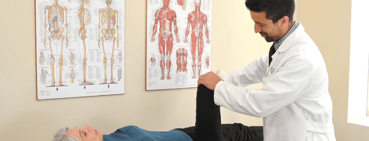
Treatment for rheumatoid arthritis may include physical therapy and low impact exercise to improve mobility, anti-inflammatory medications to alleviate pain, steroids, corticosteroids to reduce inflammation, disease-modifying antirheumatic drugs (DMARDs) or biologics to slow the progression, and joint replacement surgery. There is no cure for RA, but the goal of treatment is to limit joint damage and put the disease into remission.

What is Osteoarthritis?

Osteoarthritis, on the other hand, is a degenerative joint disease that primarily affects the cartilage–the tissue that covers the ends of the bones in a joint. Over time, the cartilage breaks down, causing bones to rub against each other. This leads to pain, swelling, and decreased mobility. Unlike RA, OA is generally associated with wear and tear of the joints. Symptoms, including pain, tenderness, stiffness, and loss of flexibility, are typically localized to the affected joints. Risk factors for osteoarthritis include age, joint injuries, repetitive stress on the joint, and obesity.

Atlantic Orthopaedics’ Dr. Akhil Sastry is a board-certified orthopedic surgeon who specializes in hip and knee joint replacement. He is a pioneer of robotic-assisted total knee replacement and has performed the surgery over 1,000 times. A recent patient had this to say about his experience with Dr. Sastry:

“I had an excellent experience and result with Dr. Sastry. He worked with me prior to surgery to ensure I could continue the activities important to me despite having osteoarthritis in the knee. After a year and a half, we agreed that I needed surgery and Dr. Sastry gave me a partial knee replacement. The pre op communication and approach were not only effective but used the latest practices to minimize pain and recover quickly. The entire surgical team helped make the surgery itself a success. But the proof is in the result, after 8 weeks of physical therapy, I am back doing the sports I love like tennis and bike riding. I am very grateful to Dr. Sastry and his team.”

Below, Dr. Sastry answers a frequently asked question about osteoarthritis. 

What are the best ways to handle osteoarthritis in my knee and what are the options for treatment for a very active senior who wants to stay active?*

Treatment options range from weight loss, low impact exercises (biking, elliptical, swimming), over the counter anti-inflammatory medications, injections, and joint replacement surgeries. Depending on the severity of disease and the limitations that are inflicted, an orthopedic surgeon can implement a treatment program that would be the most suitable for your needs.

Managing Osteoarthritis in the Knee: Treatment Options for Active Seniors

  • Physical activity: Staying active is one of the most effective ways to manage knee osteoarthritis. Gentle exercise like walking or swimming can help maintain joint flexibility and strengthen the muscles around the knee, providing better support and joint stability.
  • Weight management: Maintaining a healthy weight reduces the stress on your knees. Even a small amount of weight loss can significantly decrease the load on your knee joints, alleviating pain and slowing the progression of OA. A diet rich in anti-inflammatory foods like fruits, vegetables, and omega-3 fatty acids can also help improve symptoms.
  • Physical therapy: A physical therapist can create a personalized exercise program to improve your knee’s strength and flexibility. They can also teach you techniques to modify your movements to reduce pain and prevent further damage.
  • Injections: Corticosteroid injections can provide temporary pain relief by reducing inflammation in the knee joint. Hyaluronic acid injections, which mimic the natural fluid in your knee, can also help lubricate the joint.
  • Surgical options: If conservative treatments aren’t providing sufficient relief and your mobility is impaired, surgery may be advised. Arthroscopy, a minimally invasive procedure, can be used to remove damaged cartilage or bone fragments. In more severe cases, partial or total knee replacement surgery might be necessary. These procedures can significantly reduce pain and improve function, allowing you to remain active.

If you are suffering from joint pain due to arthritis, don’t delay getting treatment. There are many options, both non-surgical and surgical, that can improve your quality of life and get you back to doing the activities you love. Schedule a consultation with Dr. Sastry to discuss a treatment plan tailored to your needs and lifestyle.

*Medical Disclosure: The information provided on this blog is for educational and informational purposes only. While we strive to provide accurate and up-to-date information, we do not dispense medical advice or treatments to individuals who have not been seen by a healthcare professional.

It’s crucial to understand that every individual’s medical situation is unique, and what may work for one person may not necessarily work for another. Additionally, individuals may have underlying health conditions, allergies, or other factors that require personalized attention and consultation with a qualified healthcare provider.

Therefore, we strongly advise individuals to consult with a licensed healthcare professional before initiating any new treatments, making changes to their current treatment regimen, or addressing any medical concerns. This includes seeking professional guidance for managing allergies, assessing potential medication interactions, and ensuring overall safety and efficacy of any suggested treatments.

We are not liable for any actions taken based on the information provided on this blog. The responsibility for healthcare decisions lies solely with the individual and their healthcare provider. If you have any questions or concerns regarding your health, please consult a qualified healthcare professional promptly.

Exactech Computer-Assisted Shoulder Replacement

Patients suffering from severe shoulder pain know that it can impact every aspect of life, from mobility to sleep to the ability to perform basic daily tasks like getting dressed or pushing a grocery cart. The shoulder is a ball and socket joint, comparable to the hip, and has a greater range of motion than any other joint. It’s made up of three bones: the humerus (upper arm), scapula (shoulder blade), and clavicle (collarbone), which converge at the socket. The head of the humerus is the “ball” that fits into the glenoid cavity, the “socket” part of the scapula, and is stabilized by a ring of cartilage called the glenoid labrum. Damage to this protective cartilage can cause the bones to rub together, leading to joint pain that can be debilitating.

Damage to the shoulder joint can occur as a result of osteoarthritis, rheumatoid arthritis, rotator cuff tear arthropathy, or bone deterioration caused by restricted blood (avascular necrosis), among other conditions. For patients who haven’t found relief with non-surgical treatments like physical therapy or steroid injections, shoulder replacement surgery may be recommended. The outpatient procedure involves making an incision to access the shoulder joint. The surgeon then removes the damaged cartilage or parts of bone, and replaces them with metal and/or plastic implants.

X-rays have traditionally been used to assess a patient’s shoulder before replacement surgery, but x-ray doesn’t provide a comprehensive view of the joint. Now, some orthopedic surgeons–including our own Dr. Noerdlinger–have embraced a state-of-the-art technology called Exactech Guided Personalized Surgery that creates a detailed 3D model of the shoulder joint. The visual map allows the surgeon to plan and virtually simulate the surgery in advance, and utilize computer-assisted guidance during the procedure. The replacement surgery can be tailored according to the individual patient’s unique anatomy, allowing for optimal implant placement. Exactech GPS enhances precision and accuracy, resulting in better functionality, fewer complications, and an expedited recovery process.

Specializing in shoulder surgery (both Arthroscopic & Shoulder Replacements), our own Dr. Noerdlinger has been voted Top Orthopedic Surgeon by New Hampshire Magazine every year from 2017 to 2023. Dr. Noerdlinger performed 140 replacement surgeries in 2023 and is recognized as one of the top shoulder surgeons in New England. Below, he answers some questions about Exactech GPS.

In your own words, how does this procedure benefit your patients?
The Exactech GPS system allows surgeons to place the shoulder replacement in the most accurate position, which improves outcomes.

Who is a good candidate for Exactech Guided Personalized Surgery?
Anyone considering a shoulder replacement.

Where do you perform this procedure? 
I perform the surgery at NECOS and at York Hospital.  

Anything else you’d like current or future patients to know about this procedure and how it would benefit them?
Patients considering a shoulder replacement should know that it’s an outpatient procedure and the recovery is quicker than many think. Patients often don’t need post-operative physical therapy. For a Reverse Total Shoulder Replacement, patients can expect to wear a sling for one week, and patients undergoing Anatomic Total Shoulder Replacement can expect to wear a sling for about 6 weeks.   

If you’re suffering from shoulder pain and reduced joint function, schedule a consultation with Dr. Noerdlinger to find out if you’re a candidate for Exactech GPS-assisted shoulder replacement surgery. Don’t postpone getting help, so you can get back to enjoying the activities you love.

Relief From Hip Pain – How Arthroscopy Can Alleviate Pain and Restore Function

Hip pain can be debilitating, affecting everything from mobility to sleep to overall quality of life. The hip joint is where the thigh bone (femur) connects to the pelvis and is used constantly when one walks, bends, and supports his/her upper body. 

Who is a good candidate for Hip Arthroscopy?

Hip pain has varied causes; in young people the most common cause  is a labral tear–damage to the soft tissue lining the inside of the hip socket (acetabulum). A Labral tear can occur from repetitive wear over time, impingement from an underlying problem with the joint structure, or trauma from a sports injury or accident.  

For patients who suffer from a labral tear –and who have not found relief from conservative treatments like rest, over-the-counter pain relievers and physical therapy–hip arthroscopy may be the answer. It’s a minimally invasive procedure that can restore hip function and alleviate pain.

Patients with osteoarthritis, cysts, or ankylosis (fusion) of the hip joint are not good candidates for hip arthroscopy.  

What is involved in the Hip Arthroscopy procedure?

First, an orthopedic specialist will determine whether hip arthroscopy is appropriate based on a physical exam and x-ray or MRI imaging. Unlike open surgery, arthroscopy only requires a small incision through which a camera (arthroscope) is inserted. This gives the surgeon a clear, magnified view of the interior of the hip joint during the procedure. The surgeon will then repair or remove the damaged labrum, bone impingement, or loose bodies. Performed in  an outpatient setting , arthroscopic hip surgery usually leads to less post-operative pain and shorter recovery time than open hip surgery. 

Surgical Expertise + State of the Art Technology

Dr. Tyler Welch is a board certified orthopedic surgeon who has performed approximately 300 hip arthroscopy procedures – In addition to his extensive experience, Dr. Welch utilizes state-of-the-art technology, including “HipMap” software that generates a detailed 3D image of the hip structure in advance of the procedure. Used as a planning tool, this advanced imaging enables him to precisely target the surgery and minimize disruption to surrounding tissue. 

Another innovative device employed by Dr. Welch is the Guardian, a cutting-edge operating table designed to hold the patient comfortably and securely in place, while providing the surgeon greater range of motion and visibility during the procedure. Traditionally, achieving proper positioning for hip surgery required the use of a post between the patient’s legs, a technique  that can cause groin pain . The Guardian table has eliminated post-operative groin pain. 

Below, we asked Dr. Welch a few questions about hip arthroscopy.

Where do you perform this procedure?
I perform hip arthroscopy at NECOS and at York Hospital.

Are you still the only practice in the Seacoast that uses the Guardian Table for this procedure?
That’s right, I’m still the only surgeon on the Seacoast that uses the Guardian Table.

Anything else you’d like current or future patients to know about this procedure and how it would benefit them?
What is nice about hip arthroscopy is that it is a relatively straightforward recovery.  Crutches are only required for about one week, and most patients make significant gains within 4-6 weeks. It is not too painful, and I have found that most patients only require pain medication for the first 24-48 hours.

Also, surgery is reliable for the right patient.  As long as the patient has hip impingement without any sign of arthritis, surgery typically alleviates pain and eliminates most symptoms.

If you suffer from hip pain, don’t ignore it. Atlantic Orthopaedics and Sports Medicine offers treatments–both surgical and non-surgical–that can alleviate hip pain and restore your quality of life. If you think that hip arthroscopy may be right for you, schedule a consultation with Dr. Welch to learn more.

A man standing in front of a robotic total joint replacement machine at a medical conference.

Embracing Cutting-Edge Technology: Dr. Sastry’s Experience with Stryker at the AAOS Conference

As orthopedic surgeons, staying at the forefront of advancements in medical technology is crucial for providing the best possible care to patients. Recently, Dr. Sastry had an exciting opportunity that not only showcases the innovative strides in orthopedics but also highlights the trust in his skills. Stryker, a leading medical device company in orthopedics, reached out to Dr. Sastry after learning about his plans to attend the 2024 American Academy of Orthopaedic Surgeons (AAOS) conference. The request? To conduct a live demonstration of the robotic-assisted total knee replacement on Thursday, February 15th. This wasn’t his first time being asked to showcase this groundbreaking procedure; he had the honor a few years ago as well.

The AAOS Conference
The AAOS conference is the largest orthopedic gathering nationally, attracting thousands of surgeons from around the world. This prestigious event was hosted in San Francisco, providing a platform for professionals to exchange knowledge, discuss advancements, and explore the latest technologies in the field. It’s an incredible opportunity to network, learn, and contribute to the ever-evolving landscape of orthopedic surgery.

Stryker’s Leadership in Orthopedics
Stryker’s invitation underscores their position as a trailblazer in the orthopedic industry. With a focus on developing cutting-edge medical devices, Stryker has consistently pushed the boundaries of innovation. Their commitment to advancing technology has not only transformed surgical procedures but has also improved patient outcomes, making them a trusted name in the medical community.

Robotic-Assisted Total Knee Replacement
The robotic-assisted total knee replacement is a prime example of the revolutionary technologies introduced by companies like Stryker. This procedure combines the precision of robotics with the expertise of surgeons, enhancing accuracy and optimizing the patient’s experience. As Dr. Sastry demonstrated this technique at the AAOS conference, it’s a testament to the collaborative efforts between medical professionals and industry leaders to bring about positive change in orthopedic care.

Promoting the Future of Orthopedics
The significance of this opportunity extends beyond the conference walls. It’s an opportunity to showcase the advancements in orthopedics to a global audience, but more importantly, it’s an opportunity to promote the benefits of such groundbreaking procedures offered to our patients. By embracing innovative technologies, Atlantic Orthopaedics & Sports Medicine can offer patients improved outcomes, quicker recoveries, and a better quality of life.

Dr. Sastry enjoyed sharing this experience with colleagues from around the world and, more importantly, with patients who stand to benefit from these advancements in orthopedic care.

Relief From Lower Back Pain With SI Fusion

Living with chronic lower back pain can have a profound impact on not just your physical well-being, but your overall quality of life. Many people suffer from back pain due to dysfunction in the sacroiliac (SI) joint, which connects the bottom of the spine (sacrum) to the pelvis (ilium). These two joints, one on each side act as a shock absorber for the spine and play a crucial role in stability and weight bearing ability, transferring pressure from the lower back to the legs. The SI joint can be damaged by traumatic injury, a degenerative condition, or tissue loosening from things such as connective tissue disorders and pregnancy. Patients with SI dysfunction will feel pain in the low back, especially when walking, standing up from a seated position, or lifting. Other symptoms can include numbness or tingling in the lower extremities; pain in the pelvis, hip, or groin; pain radiating down the legs; or buckling in the legs.

Diagnosing SI Dysfunction 

Because low back pain can be a symptom of other conditions like sciatica, diagnosis is usually done with a physical exam, followed by an injection that will temporarily block the pain if it’s originating in the sacroiliac joint. In some cases, the pain can be managed with anti-inflammatory medications, physical therapy, or steroid injections, but for patients who haven’t responded to those treatments there is a minimally invasive procedure called SI Fusion that may offer sustained relief. 

What is the SI Fusion Procedure?

SI Fusion, short for sacroiliac joint fusion, is a minimally invasive procedure in which the surgeon inserts an implant into the SI joint space. Guided by real-time imaging, a small incision is made and the implant is fixed in place, fusing the sacrum to the ilium in order to limit movement. This fusion, by restricting excessive motion in the SI joint, restores stability and alleviates pain. 

What Does the Recovery From SI Fusion Involve?

Because the procedure is minimally invasive, the risk of complications is low and recovery times are quicker than with traditional open surgery. Patients can expect to be mobile immediately after the procedure, and most experience rapid pain relief. Post-operative physical therapy may be recommended to restore function in the joint and aid recovery.

Below, a Q&A about SI Fusion with pain management specialist Dr. Lehn:

How does this procedure benefit your patients?

15-30% of low back pain comes from the SI joint. The SI joint can be a difficult pain to treat as many times PT, medications, and injections only provide temporary relief.

Who is a good candidate for an SI fusion?

If a patient has 90% relief of pain from a SI injection and the pain keeps returning despite exercise, medications, and repeated injections, then they can be considered for SI fusion.

Where do you perform this procedure?

The procedure is performed in the operating room but is done with minimal sedation, and the patient can walk out of the recovery room and go home the same day as the procedure.

Do you have a recent patient success story to share? 

One patient had her left side done in June and felt so good she came back in September to have the other side done! She is very happy and back to her very active lifestyle.

Anything else you’d like current or future patients to know about this procedure and how it could benefit them? 

This is a very minimally invasive procedure with minimal downtime. The pain from the procedure is minimal. Unfortunately, intense activity is limited for 6 weeks but patients are up and walking the same day. Fusions sound really big and dangerous, but it is essentially a stabilization procedure. SI joints are often painful due to excessive movement. The SI stabilization procedure takes away the additional movement and helps return the patient to a normal functioning joint.For patients grappling with chronic lower back pain caused by SI dysfunction, the SI Fusion procedure can provide long-term pain relief and renewed quality of life. If you suffer from pain that hasn’t responded to non-surgical treatments, schedule a consultation with Dr. Lehn to explore whether SI Fusion is right for you. Take your first steps toward a pain-free future!

Knee Pain From a Dislocated Kneecap? 

Patella Instability Surgery Can Provide Relief!

What Is Patellar Instability?

When the kneecap shifts out of its normal alignment, you may feel pain, trouble walking, a buckling or locking sensation, or a feeling of the knee “giving way.” Patella instability can impact daily life and make it difficult to stay active. 

How Does the Patella Instability Procedure Work?

In this procedure, the surgeon carefully realigns the patella within the femoral groove to ensure proper tracking during knee movement. The specific surgical technique can vary based on factors like the patient’s age, and the severity of the injury. In the Medial Patellofemoral Ligament (MPFL) Reconstruction procedure that Dr. Welch describes below, the patient’s ligament was reconstructed to stabilize the patella on the inside of the knee. In cases of severe instability, the surgeon may also reposition the tibial tubercle–the bony bump below the patella–to improve patellar tracking.

What Can I Expect Post-Surgery?

Patients can expect to go home the same day as the procedure. Recovery time varies and patients will need to use crutches and wear a knee brace for a period of time, typically 4-6 weeks. Physical therapy is also an important part of the recovery process.

We sat down for a Q&A with our Orthopedic Surgeon and Sports Medicine Specialist Dr. Welch. 

In your own words, how does this procedure benefit your patients? 

Patella instability is a condition in which the patella (kneecap) dislocates from the femur (thigh bone). Most patients are young, often teenagers. Fortunately we can fix this condition surgically. This procedure benefits patients because it generally restores one’s confidence and his/her ability to play sports.

Who is a good candidate for the Patella Instability Procedure?

A good candidate for patella instability surgery is anyone who has had at least one patella dislocation or multiple subluxations (partial dislocations).

Where do you perform this procedure? 

I perform this procedure at New England Center for Orthopedic Surgery (NECOS) in Portsmouth and at York Hospital in York, Maine. 

Do you have a recent patient success story you can share?

Recently, a 15 year old male high school student came to see me with his family. The patient had sustained two dislocations prior to his visit with me. These dislocation events are painful and stressful, and the young man was apprehensive about playing sports ever again unless we fixed his knee. He underwent a procedure–a medial patella femoral ligament (MPFL) reconstruction–to stabilize his patella. Surgery was straightforward and he recovered nicely. He required crutches for 10 days and a brace for four weeks. After six weeks, he had normal motion and no swelling in his knee. I cleared him to return to sports three months after surgery.

Anything else you’d like current or future patients to know about this procedure and how it would benefit them?

There are a few important things to consider regarding patella instability surgery. The procedure is very reliable – dozens of studies have demonstrated success rates in the 90-95% range, and my experience has been similar. The vast majority of these patients are happy because they regain “trust” in their respective knees, and recovery is straightforward. Further, patients usually return to the same level of activity that they had prior to the dislocation(s). 

If you have knee pain from dislocation, visit our website to learn more about Arthroscopic Knee Surgery or schedule a consultation with Dr. Welch.

Is Pain in Your Thumb Holding You Back?

CMC Arthroplasty As A Solution For Thumb Pain

If you’re experiencing pain and stiffness at the base of your thumb, you might be dealing with CMC (carpometacarpal) arthritis. But don’t worry – you’re not alone. There are effective treatment options available for this common thumb condition that can provide much-needed pain relief and restore motion.

What are the symptoms of CMC arthritis?

Common symptoms include pain at the base of the thumb, especially during pinch and grip activities. The joint also may become swollen and tender to the touch. Typically, CMC arthritis decreases mobility in the thumb, and weakness may develop that makes it difficult to grasp objects.

What causes CMC arthritis?

CMC arthritis is a degenerative condition, often attributed to the aging process. Other contributing factors can include:

  • Repetitive use: activities that require repetitive thumb movements can accelerate joint wear and tear.
  • Trauma: a previous thumb injury can increase the risk of developing CMC arthritis.
  • Genetics: genetic history may predispose some people to joint degeneration.
  • Inflammatory conditions: certain immune or inflammatory conditions can contribute to CMC arthritis.

Atlantic Orthopaedics’ hand surgeon Dr. Meyer answers some common questions about CMC Arthroplasty, a life-changing procedure for patients suffering from advanced thumb arthritis.

In your own words, how does this procedure benefit your patients?
Arthritis at the base of the thumb (CMC arthritis) is a common source of pain and disability for adults, affecting one in three women and one in eight men over the course of their lifetime. A variety of treatment options exist, starting with conservative management (bracing, therapy, anti-inflammatories, corticosteroid injections) and progressing to surgery. For advanced thumb CMC arthritis that does not respond to the above treatments, CMC arthroplasty can provide lasting pain relief and improvement in thumb strength and motion.

There are a variety of surgical techniques for CMC arthroplasty. Most involve the removal of the painful, arthritic bone at the base of the thumb (trapezium) and use of a nearby tendon to stabilize the thumb in its native position. Recent innovations in the field of hand surgery have allowed for the use of a non-absorbable suture instead of a patient’s own tendon to maintain the thumb’s resting position. This procedure, termed a suture-button suspensionplasty, avoids complications from harvesting nearby tendons and allows for accelerated rehabilitation and recovery after surgery. While most other techniques require between four to six weeks of immobilization in a cast, I allow my patients to start gentle motion after their first post-operative visit (approximately 10 days after surgery) when using this technique.  

Who is a good candidate for this procedure?
Thumb CMC arthritis can affect anyone, but it becomes more prevalent with older age. This procedure is typically indicated for middle-age and elderly adults who have painful arthritis at the base of their thumb that limits their daily activities.

Where do you perform this procedure ?
I perform thumb CMC arthroplasty procedures at NECOS, Portsmouth Regional Hospital, and York Hospital. 

Do you have a recent patient success story you can share?
A 65-year-old retired professional pianist presented to the office with painful thumb CMC arthritis that failed to improve with bracing and steroid injections. Alleviating pain and preserving thumb motion were her biggest priorities. Due to her activity level, we elected to perform a CMC arthroplasty using the suture-button suspensionplasty technique.

Her surgery and post-operative course both progressed very smoothly. She was seen at her first post-operative visit and transitioned to a removable brace that allowed for gentle motion. At six weeks, she began gentle strengthening under the guidance of our occupational therapy team. At three months, she was cleared to return to full activity. She is back to playing piano and feels that her motion is even better than before surgery because she is no longer limited by pain. We were thrilled to help her get back to one of her favorite activities and to enable her to keep teaching other aspiring pianists.   Anything else you’d like current or future patients to know about this procedure and how it would benefit them?
Thumb CMC arthroplasty yields excellent results, but it is not the only treatment recommendation for patients with thumb CMC arthritis. There are a variety of non-surgical and surgical treatments that can help with pain at the base of the thumb prior to considering a CMC arthroplasty. Every patient evaluation begins with a thorough clinical history, physical examination, and radiographs (x-rays) of the hand. From there, I work with each patient to understand their goals before we collectively decide on an appropriate treatment plan moving forward.

CMC arthritis can be a debilitating condition, but there is help available. For many patients, CMC Arthroplasty has proven to be an effective way to alleviate pain and restore quality of life. If you’re suffering from pain and limited function in your thumb, schedule a consultation with Dr. Meyer to discuss whether CMC Arthroplasty may be right for you.

Pain Relief for Rotator Cuff Tears–Introducing the Stryker InSpace Balloon

What is a Rotator Cuff Tear?

The rotator cuff is a group of four tendons that surround and stabilize the shoulder joint, connecting the muscles of the upper arm to the shoulder blade. When a rotator cuff tear occurs–from acute trauma, repetitive stress, or degeneration over time–the tendon becomes separated from its attachment point on the bone. Rotator cuff tears are painful and cause weakness in the joint and limited shoulder mobility. Common symptoms of a tear include difficulty lifting your arm, a popping or crackling sensation in the shoulder, and pain both at rest and with movement.

Unfortunately, rotator cuff tears don’t heal on their own. If an untreated tear results in arthritis in the joint, patients often require shoulder replacement surgery, but for patients who still have shoulder function and haven’t developed arthritis, there is a less invasive treatment option called the Stryker InSpace Balloon.

What is the Stryker InSpace Balloon?

The Stryker InSpace Balloon is a medical device used to treat rotator cuff tears. It’s made of biocompatible material–a substance that can be implanted in the body and tolerated without damaging existing tissue. The balloon creates more space within the subacromial area–the space between the bone at the top of the shoulder and the ball of the shoulder–reducing compression and friction caused by the torn rotator cuff.

What’s involved in the procedure?

The Stryker InSpace Balloon is placed arthroscopically, using a small incision and the guidance of a camera to precisely place the deflated balloon in the subacromial space. The balloon is then inflated with a sterile saline solution, causing it to push the acromion away from the rotator cuff tendons. This reduces impingement on the injured tissues to restore function and alleviate pain.

The procedure is minimally invasive and performed on an outpatient basis. Recovery time varies, depending on the individual patient and the extent of the injury, and rehabilitation may include physical therapy to help restore strength and mobility in the shoulder.

How do I know if the Stryker InSpace Balloon can help me?  

If you’re experiencing shoulder pain, or have been diagnosed with a rotator cuff tear, it’s important to consult with a shoulder specialist as soon as possible. The specialist will evaluate your injury and medical history, and may recommend the Stryker InSpace Balloon procedure as a treatment option.

Below is our Q&A with Dr. Noerdlinger, an orthopedic surgeon specializing in the shoulder. Dr. Noerdlinger is a partner at Atlantic Orthopaedics with extensive experience performing the Stryker InSpace Balloon procedure.

Q: In your own words, how does this procedure benefit your patients? 
A: This is a pain relieving procedure.

Q: Who is a good candidate for the Stryker InSpace Balloon procedure?
A: The procedure is indicated for patients with irreparable rotator cuff tears, with good shoulder function and minimal to no arthritis.

Q: Where do you perform this procedure? 
A: I perform the procedure at NECOS (New England Center for Orthopaedic Surgery) in Portsmouth, and York Hospital in Maine.

Q: Do you have a recent patient success story you can share?
A: I performed the Stryker InSpace Balloon procedure on a 67 year old man with years of pain and an unfixable rotator cuff tear. He described his shoulder as getting worse despite physical therapy and injections. He had pain with an active range of motion, and stated his shoulder was 30% normal. Four months after his February surgery, he described his pain as a 2 out of 10 on a daily basis and rated his shoulder as 60% normal.

I also treated a 73 year old female patient who came to me with pain from an irreparable rotator cuff tear. She described her shoulder as 40% normal before the procedure and 70% normal 3 months after her March 1st surgery.

Another patient, a 60 year old man with bilateral irreparable rotator cuff tears, had the balloon placed in his right shoulder on March 1st, and his shoulder improved from 20% normal to 65% normal in 6 weeks. He then underwent left shoulder balloon placement May 10th.

Q: Anything else you’d like current or future patients to know about this procedure and how it would benefit them?
A: The rotator cuff is the one structure that, when torn, causes the most pain and dysfunction in the shoulder. Rotator cuff tears do not heal, get bigger over time, become unfixable, and cause arthritis. Rotator cuff repair surgery is very successful and can restore normal, pain-free function.

If the rotator cuff is irreparable, the painful shoulder is often treated with a reverse shoulder replacement. For patients with unfixable, painful rotator cuff tears with little to no arthritis and good function, an InSpace balloon is a good option. 

If you’re suffering from shoulder pain from a rotator cuff tear that hasn’t responded to treatments like injections or physical therapy, schedule an appointment to find out if the Stryker InSpace Balloon procedure is right for you.

Torn Meniscus? Eliminate Knee Pain With This Procedure 

The meniscus is a C-shaped piece of cartilage in the knee joint that plays a crucial role in function and stability. The two meniscii in each knee function as shock absorbers. They are composed of dense fibrocartilage, which is more flexible than bone but firmer than other types of cartilage. Meniscus pads play an important role in walking, running and jumping, and they aid in weight distribution and alignment. The meniscus also helps keep the knee joint lubricated to reduce friction between the femur (thigh bone) and tibia (shin bone). 

A meniscus root tear is a relatively common knee injury that occurs when the meniscus separates from its attachment point on the tibia bone. This type of tear can be caused by the wear and tear that happens with age, or by trauma or injury, such as a sports injury. People who suffer a tear often feel a “popping” sensation in the back of the knee, followed by pain, instability, and a loss of function. Untreated, a meniscus root tear can lead to osteoarthritis and degeneration in the knee. Fortunately, there is a minimally invasive procedure available to fix the issue.

Atlantic Orthopaedics’ surgeon Dr. Welch specializes in meniscus root repair, having performed more than 100 successful procedures to restore function, eliminate pain, and prevent further damage. In this surgery, he makes three small incisions, prepares the torn end of the meniscus for repair, then securely reattaches the meniscus to the root attachment site on the tibia bone through a small tunnel in the tibia. 

Following surgery, Dr. Welch places the patient’s knee in a brace to protect the repaired meniscus and allow it to heal. Physical therapy is an important part of the recovery process. Recovery from meniscus root repair takes time and it’s important to follow your surgeon’s instructions regarding weight-bearing and the use of crutches. It may take three months for the knee to fully heal, but the vast majority of patients have excellent outcomes and experience a return to normal knee function.

Below, Dr. Welch answers some questions about Meniscus Root Repair:

Q. In your own words, how does this procedure benefit your patients suffering from knee pain

A. A meniscus root tear is a specific type of meniscus tear that is more serious than a typical meniscus tear. If the root tears, the meniscus becomes loose and unstable. Surgery involves repairing the meniscus back to the tibia bone. This procedure benefits patients because it restores the function of the meniscus, eliminates pain, and helps restore patient confidence in his/her knee.

Q. Who is a good candidate for a Meniscus Root Repair? 

A. A proper candidate for meniscus root repair is a patient who sustains a root tear who does not have significant arthritis in the knee.

Q. Where do you perform Meniscus Root Repairs ? 

A. I perform meniscus root repairs at New England Center for Orthopedic Surgery (NECOS) in Portsmouth, NH and at York Hospital in York, ME.

Q. Do you have a recent patient success story you can share?

A. I have dozens of patient success stories. Meniscus root repairs are very reliable; as long as the meniscus heals to the tibia, patients usually return to normal function with no issues. Previous studies have revealed success rates > 90%.

Q. Anything else you’d like patients to know about this procedure? 

A. I have many patients (>100) who have had tremendous success after a meniscus root repair. Unfortunately, one issue with this procedure is that each patient must use crutches or a walker for the first 5-6 weeks after surgery. This is inconvenient, but necessary, so that the meniscus has time to heal.

If you’re suffering from knee pain and loss of mobility from a meniscus root tear, you may be a good candidate for this minimally invasive procedure. Schedule an appointment with Dr. Welch for an evaluation: https://www.atlanticorthopaedics.org/providers/tyler-welch-md/

Running person icon to the left of the image and to the right is the blog title: What is Collagen Augmented Rotator Cuff Repair?

What is Collagen Augmented Rotator Cuff Repair?

Rotator cuff injuries are one of the most common causes of shoulder pain, affecting more than 2 million Americans each year. If you have a partially torn rotator cuff that hasn’t responded to non-surgical treatment like physical therapy and cortisone injections, you may be a candidate for a procedure called Collagen Augmented Rotator Cuff Repair, performed by our shoulder specialist Dr. Noerdlinger.

What is the rotator cuff?
Shoulders are not singular joints, but rather a complex arrangement of bones, cartilage, ligaments, muscles, and tendons. The rotator cuff is made up of four muscles, connected by tendons, that originate from the shoulder blade (scapula) and attach to the upper arm bone (humerus) to form a cover, or “cuff.” The rotator cuff provides stability to the shoulder joint and helps the arm lift and rotate. 

What causes a rotator cuff tear?
A rotator cuff tear happens when the tendons pull away from the bone. With a partial tear, the tendon remains attached to the bone and with a full tear it separates from the bone completely. Tears most commonly happen as a result of normal wear and tear due to aging, with people over 40 at greatest risk. A tear can also be caused by an acute injury like a fall, from repetitive shoulder movement from jobs like carpentry or painting, or from sports like tennis or rowing. Untreated, a rotator cuff tear can worsen over time and may lead to chronic shoulder pain and limited range of motion.

How do I know if I have a rotator cuff tear?
The most common symptom of a rotator cuff tear is pain. A tear can also cause weakness in the arm and shoulder, difficulty raising your arm or lifting something, and a clicking or popping sensation in the shoulder. You may also experience swelling or tenderness in the shoulder joint. If you think you have a rotator cuff tear, a doctor will need to perform a physical exam and may confirm your diagnosis with an x-ray, MRI, or ultrasound.

What is Collagen Augmented Rotator Cuff Repair?
Collagen augmented rotator cuff repair is a surgical technique that uses a collagen implant to reinforce the rotator cuff repair. The implant is made of a biologically derived material similar to the collagen found in human tissue. It’s designed to provide additional strength and support to the healing tendon, reducing the risk of re-tear. The postage-size collagen patches are absorbed by the body within about 6 months, and patients generally experience shorter recovery times than with conventional rotator cuff surgery.

We recently sat down with shoulder specialist Dr. Noerdlinger and asked him a few questions about this procedure. 

Q: How does this procedure benefit your patients?
A: Collagen implants can lead to quicker recoveries. Adding collagen facilitates the biologic healing of torn tendons in certain situations. They are used in two situations:

  • To repair a partially torn rotator cuff tendon. In this scenario, the collagen is laid on top of the partially torn tendon. The implant blends in with the thinned rotator cuff tendon, thickens it and allows it to heal. Patients, with partial tears, do not need to wear a sling after surgery.
  • To augment a repaired shredded tendon that may not heal on its own, particularly in the setting of a revision rotator cuff tear when the tendon did not heal after the first surgery. 

Q: Who is a good candidate for the Collagen Augmented Rotator Cuff Repair Procedure?
A: Not everyone is a candidate for this procedure. Patients with irreparable rotator tears would not qualify for this procedure. Also, patients with good tissue may not need to augment their repairs. 

Q: Where do you perform the Collagen Augmented Rotator Cuff Repair procedure?
A: The procedure requires anesthesia and is performed arthroscopically at NECOS and York Hospital.

Q: Can you tell us about a success story? 
A: I recently saw a 56 year old female with worsening constant shoulder pain since April 2022. I performed a Partial Rotator Cuff Repair with collagen implant in January of 2023. She was seen in early March of 2023 and reported to be doing well with minimal complaints of pain. 

NOTE: Shoulder surgery has a long recovery and often people take 4-6 months before they feel great. Having an expert shoulder surgeon like Dr. Noerdlinger may help speed up recovery. 

AOSM Running Man logo and Blog Title "New Pain Management Procedure"

Introducing MILD: life-changing relief from Spinal Stenosis

Does spinal stenosis make it painful for you to stand or walk for more than a few minutes? We’d like to tell you about a safe and effective treatment option called MILD, or minimally invasive lumbar decompression, offered by our pain management specialist Dr. Lehn

What is spinal stenosis?

Inside the spine is a hollow channel called the spinal canal that contains the spinal cord and nerves. Spinal stenosis occurs when the channel narrows, putting pressure on the spinal cord and nerves. In addition to pain, symptoms may also include weakness, numbness, and cramping in the lower back and legs. It’s most often caused by the normal wear and tear that comes with aging, but factors like arthritis and protruded discs can also lead to spinal stenosis.

How is it treated?

Spinal stenosis is often a progressing problem and it is difficult to predict if a patient will ultimately return to normal once symptoms begin. Many patients will benefit from treatments like physical therapy or steroid injections but the duration of benefit varies patient to patient. Some people manage the problem with minimal treatment while others need surgical decompression to relieve the pressure on the spinal cord. Understandably, many patients are hesitant to undergo invasive surgery, and may be worried about a hospital stay and a lengthy recovery time. 

What is MILD?

The MILD procedure for spinal stenosis is a minimally invasive alternative to traditional spinal surgery that has provided immediate relief and life-changing results for many patients. Guided by x-ray, Dr. Lehn inserts a spinal decompression device through a tiny incision in the back, using it to remove a portion of the tissue that’s pressing on the spinal nerves. The procedure takes less than an hour and, unlike traditional surgery, MILD is performed with local anesthesia and sedation, so you can go home the same day. Patients who undergo MILD typically experience minimal pain and much faster recovery times than with traditional surgery.

We sat down for a Q&A with Atlantic Orthopaedics’ Pain Management Specialist Dr. Lehn: 

Q: How does this new procedure benefit your patients? 

A: The MILD procedure is specifically for people with spinal stenosis diagnosed with a MRI leading to Neurogenic Claudication. Spinal stenosis is another word for a kink or tightening around the spinal nerves and Neurogenic claudication is pain that is present with standing or walking that improves with sitting. The goal of the procedure is to remove a portion of the tissue that is causing the spinal compression with a special tool using x-ray guidance. Once the patient heals they should expect a great increase in their ability to walk and stand! 

Other Benefits:

  • Almost immediate recovery from the procedure; full benefit takes 3-4 weeks.
  • 1 cm incision, no implants.
  • Minimal sedation for 30-45 minutes; walking home same day.
  • This procedure does not inhibit future surgery from being done.
  • 5-year studies show sustained relief of pain from this procedure.

Q: Do you have a recent patient success story you can share with us?

A: I just had a follow up with a 76 year old, male patient who is doing great! He was on the fence on whether he should do invasive surgery or the MILD. The patient had pain down both legs after standing and walking for just a short time.

His legs would also give out, and he had a tough time carrying things while walking. He was referred by Dr. Sutherland as he was very anxious about having a large surgical procedure so the MILD seemed like a great solution. 

He is now walking a mile with minimal pain!

Q: Who is a good candidate for the Minimally Invasive Laminectomy (MILD) Procedure? 

A: Patients who have trouble standing or walking, as long as the pain improves when they sit or lean forward on a shopping cart are good candidates for this procedure.

Q: What is the ideal age range for this procedure?

A: Any age. It is especially good for patients who do not consider themselves candidates for more invasive spinal surgery.

You might be a good candidate if: 

1. You have Spinal stenosis confirmed by MRI. 

2. You have difficulty standing and walking (but not pain lying down or sitting–that is something else.) 

3. You’ve had epidurals that work but are not long lasting.

Q: Where do you perform the MILD procedure? 

A: The procedure is performed in a surgery center. I currently perform the procedure at our state-of-the-art surgery center, NECOS.

Q. Is there anything else you’d like patients to know about this new procedure? 

A. It is very safe, with a rapid recovery, and a great option for people anxious about having invasive surgery. For the right patient it can be life changing!

If you have Spinal Stenosis and think you may be a good candidate for the MILD procedure, we invite you to schedule an appointment with Dr. Lehn for an evaluation. Why wait to get back to doing the things you love?

Atlantic Orthopaedics & Sports Medicine